
TINEA NIGRA
(Tinea Nigra Palmaris, Keratomycosis Nigricans Palmaris, Pityriasis Nigra)
Luciano Schiazza M.D.
Dermatologist
c/o InMedica - Centro Medico Polispecialistico
Largo XII Ottobre 62
cell 335.655.97.70 - office 010 5701818
www.lucianoschiazza.it
Tinea nigra is an uncommon superficial fungal infection (dermatomycosis) of the outermost layer of skin, called the stratum corneum.
It most commonly occur on the palmar aspects of hands with persistent slowly espanding demarcated dark brown to black irregularly shaped patch giving a spattered appearance that resemble silver nitrate or India ink stains.
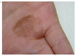
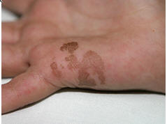
The macule appears insidiously as a small dark spot. The area tends to slowly grow over weeks to months. It has an ovoid, round, or irregular shape, and often sports a darker border. Occasionally the soles ("Tinea nigra plantaris") and, more rarely, other surfaces of the skin (neck or chest wall), can also be affected. The lesions may extend to the fingers or toes, respectively.
Lesions are non-inflammatory and slightly scaly and disease do not itch or sting in most cases, but it may be associated with pruritus. The borders are typically discrete and the pigmentary change may appear mottled or velvety. Familial spread of infection has also been reported.
Macules are typically solitary, although more than one lesion can be present, rounded or have irregular shapes. The size may range from 1 millimeter to several centimeters in diameter, depending on the duration of the infection. The dermatomycosis tends to occur in areas with an increased concentration of eccrine sweat glands and often infects those with a tendency to excessive sweating: hyperhidrosis appears to be a risk factor for this disease.
The female-to-male predilection is 3:1. Most often occurs in pediatric and adolescent populations; however, individuals of any age may be affected. This infection is usually caused by the fungus (brown mould) formerly classifed as Phaeoannellomyces werneckii, (formerly classified as Exophiala werneckii and Cladosporium werneckii) but more recently classified as Hortaea werneckii. It is classified in the family Dematiaceae, class Hyphomycetes, phylum Deuteromycota. It is a common saprophytic fungus believed to live in rotting wood, soil, compost, or sewage, humus in humid tropical and sub-tropical regions.
Other species of dematiaceous fungi, such as Stenella araguata first described and named Cladosporium castellanii in 1973 may produce a similar clinical picture.
It is a very benign condition that often causes no discomfort apart from its appearance: this often delays the patient's decision to seek medical advice.
But sometimes Tinea nigra may be mistaken for the more serious medical disorder like melanoma. Macules showing an uneven rate of spread and/or coalescence must raise the suspicion of melanocytic nevi, junctional nevi, or melanoma. Cequeira first described tinea nigra in 1891, calling the infection keratomycosis nigricans palmaris. In 1921, Horta isolated the pathogen and gave it its original name, C. werneckii.
Infection is believed to occur as a result of direct inoculation onto the skin subsequent to an injury from contact with a contamination source such as decaying vegetation, soil, sewage, wood, or compost that involves a break in the skin in the affected area.
Typically, the incubation period is 2-7 weeks, although was as long as 20 years. The pigmentary change in the skin is due to the accumulation of a melaninlike substance in the fungus. Tinea nigra is a rare condition overall. It has a World-wide distribution: although it occurs most frequently in tropical regions of Central and South America, Africa, South-East Asia and Australia, it may develop in temperate parts of the world after travel to the tropics and subtropics.
Spontaneous resolution is rare. Tinea nigra is usually treated with topical medications that can be applied to the affected area of skin, using the combination of keratolytic preparations (e.g. salicylic acid) in conjunction with topical antifungals. To reduce the chances of getting tinea nigra, because infection is believed to occur after inoculation subsequent to trauma, patients should use care when travelling in areas where tinea nigra may be contracted, avoiding contact with potentially contaminated sources material, such as soil, sewage, compost, and decaying wood.
In the diagnosis of the disease a simple microscopic examination of the skin scrapings of the affected area readily aids the distinction between tinea nigra and other serious medical disorders that result in pigmentary changes such as a junctional nevus or acral lentiginous melanoma.
Scrapings taken from the edge of the scaly lesion mounted in 10% KOH (potassium hydroxide) show pigmented brown to dark olivaceous mycelium (a group of branched filaments or hyphae) typical of Hortaea werneckii.