ERYTHROMELANOSIS FOLLICULARIS OF THE FACE
Luciano Schiazza M.D.
Dermatologist
c/o InMedica - Centro Medico Polispecialistico
Largo XII Ottobre 62
cell 335.655.97.70 - office 010 5701818
www.lucianoschiazza.it
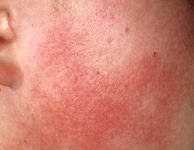
Erythromelanosis follicularis of the face (EFF), is first described in 1960 by Kitamura K. and collaborators in Japan, as a condition characterized by well-demarcated bilateral and symmetrical patches of
- Hyperpigmentation (reddish-brown pigmentation)
- erythema (with or without telangiectasia),
- follicular papules (follicular plugging)
on the cheeks and preauricular area.
EFF begins on the preauricular cheeks and gradually can spread till the submandibular areas of the neck (in this case the disease is called erythromelanosis follicularis faciei et colli – EFFC)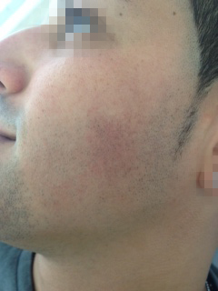
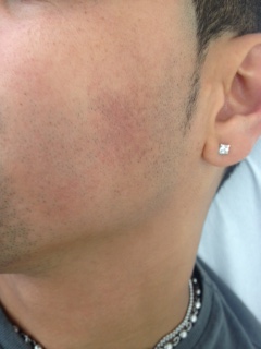
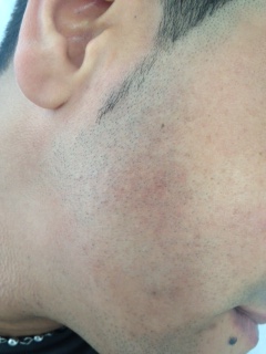
The skin texture was granular, with many, pale, follicular papules that can be appreciate as a rough surface on palpation. There was no evidence of atrophy or scarring although vellus hair appeared to be less over these areas.
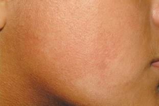
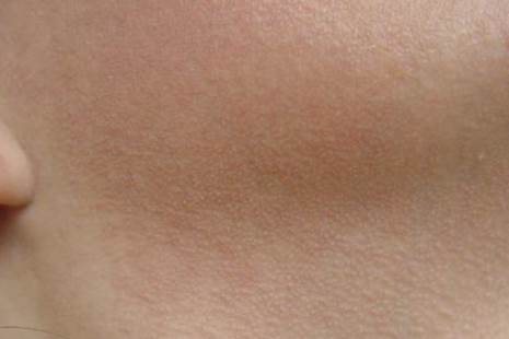
Primarily EFF is observed in adolescents. However, it also affects children and young adults, with preference for the male sex but there have been many reports of EFF also in women in the years followiong Kitamura’s description.
Bilateral distribution is the main characteristic but unilateral cases have been described.
The dermatosis is usually asymptomatic, although some patients complain of burning sensation and increased redness over the affected areas on exposure to sunlight.
Keratosis pilaris on the arms and shoulders is frequently associated with EFFC.
The disease is of unknown etiology. Some Authors consider it to be a poly-etiological disorder with the possibility of a chromosomal instability syndrome, in which an hereditary component (autosomal recessive) could interfere with its genesis.
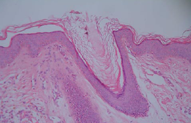
Histopathologic evaluation reveales:
-
follicular plugging
-
hyperkeratosis (thick orthokeratotic keratin layer)
-
increase pigmentation in the basal layer
-
perivascular and periadnexal inflammatory infiltrate (lymphocytic)
- dilatated blood vessels in the upper dermis
Differential diagnose (when associated with one or more components of EFFC triad) includes:
- athrophoderma vermiculatum,
- ulerythema ophryogenes
- poikiloderma of Civatte
- erythrose peribuccale pigmentare of Brocq
- keratosis pilaris rubra faciei
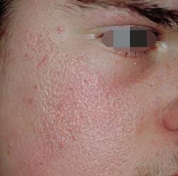
Athrophoderma Vermiculatumis a rare, benign, autosomal dominant, follicular disorder primarily affecting children with small pitted scars over cheek and forehead producing a reticulated, honeycomb worm-eaten appearance.
The individual depressions of the skin were irregular and different from pinpoint to a few millimeters in diameter. The affected area is cast with an erythematous blush.
MacKee and Cipollaro described it, with somewhat amusingly, “the cribriform pattern resembling a telescopic view of the moon at first quarter”.
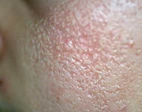
This condition was first described byUnnain 1894 as ulerythema acneiforme. Pernetin 1916 called it as atrophodermia reticulata symmetrica faciei,Mackee and Parounagianin 1918 called the condition as folliculitis ulerythematosa reticulata.Darierin 1920 coined the term atrophodermie vermiculee,Littlein 1923 used the name of folliculitis atrophicans reticulata whileWinerpreferred the term atrophoderma reticulatum.. In 1943Savarardused the descriptive name of honeycomb atrophy. It is also known as acne vermoulante, folliculitis ulerythematosa reticulata and honeycomb atrophy.
It usually begins in childhood as a symmetric worm-eaten or reticular atrophy of the cheeks that may extend to the ears or forehead. It has a slow progression. There is absence of pustules, papules, seborrhea or scaling.
Common histologic findings are: horny follicular plugs, milia, loss of rete pegs, atrophic hair follicles, sclerosis of dermal collagen, pres3ence or absence of variable degrees of perifollicular inflammation.
The primary defect in atrophoderma vermiculatum is considered to be an abnormal keratinisation in and around the pilosebaceous follicles followed by dermal atrophy. This type of atrophy is usually bilateral, but in our patient it was peculiarly unilateral.
Ulerythema(scarring with erythema)ophryogenes(related to the eyebrows) is a disease characterized by keratosis pilaris succeeded by atrophy. It mainly affects children and young adults.
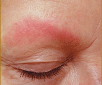
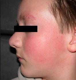
It starts during early childhood with erythema and tiny keratotic papules on the lateral parts of the eyebrows a reticular erythema and small horny papules in the outer halves of the eyebrows that may result in atrophy and permanent alopecia (loss of the involved hairs). Occasionally the disorder extends to the cheeks (Keratosis pilaris atrophicans faciei).
The affected areas may feel rough on delicate palpation.
Two stages may eventually be observed: an early stage characterized by follicular hyperkeratosis and a late stage with some evidence of fibrosis and atrophy.
Males are predominantly involved. The true cause of ulerythema ophryogenes is unknown: many cases are presumed to be the result of an inborn defect but an autosomal dominant inheritance has also been reported.
The disease often improves with age. Frequent exposure to UV radiation exacerbates ulerythema ophryogenes.
A distinction between atrophoderma vermiculatum and ulerythema ophryogenes may occasionally difficult but the latteer presents primarily on the lateral aspects of the eybrows, with erythema, follicular papules, and alopecia.
The above conditions differ from EFFC due to the presence of atrophy and scar.
Poikiloderma of Civatteis a benign condition first described by Civatte in 1923. It is characterized by symmetric patches of reddish-brown reticulated pigmentation with atrophy and teleangectasias (that spare the thin rim of skin around each follicle), on the lateral cheeks and side of the neck, except the central submental region, shaded by the chin. It is usually asymptomatic.
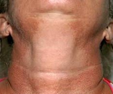
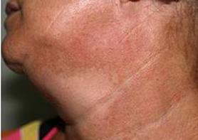
The term poikiloderma refers to the combination of atrophy, telangiectasia, and pigmentary changes (both hypopigmentation and hyperpigmentation).
Absence of follicular papules along with the starting age and lesions’ distribution, differentiate this from EFF.
In the etiology of poikiloderma of Civatte many factors may be implicated:
- Chronic UV exposure with cumulative actinic damage
- Hormonal changes related to menopause or low estrogen levels
- Genetic predisposition (autosomal dominant inheritance with variable penetrance) expressed as an increased seusceptibility to UV radiation.
- Photosensitizing chemicals in perfumes or cosmetics.

Erythrose peribuccale pigmentare of Brocq,also known erythrosis pigmentosa mediofacialis, is a disorder of the mediofacial area and is common in women.
Described for the first time by Brocq in 1923, the disease is characterized by scaling and an orange-yellow-brown to erythematous discoloration around the nose and chin (the middle of the face).
Various names were given to the disease in the following years: erythrose pigmentaire faciale, dermatose pigmentée médiofaciale, erythrosis pigmentosa peribuccalis, erythrosis pigmentata faciei, erythromelanosis follicularis faciei, erythrosis pigmentosa faciei et colli.
Keratosis Pilaris Rubra Faciei, is characterized by bright erythema and keratotic follicular papules located on the cheeks. Patients may experience a flkushing and blushing sensation. Sometimes it is misdiagnosed as rosacea.
Unilateral casesmay extend the differential diagnosis to:
- Nevus of Ota,
- Becker’s melanosis,
- Berloque dermatitis
- Fixed drug eruption
- Cafè-au-lait macules
Because of seasonal influences it should be avoided solar exposition as well as other sources of ultraviolet light and sunscreen is recommended. Various treatment options have been described sometimes satisfactory but lesions can recur soon after the treatment was discontinued: topical moisturizers, topical keratolytics, peelings. Laser can attenuate hyperpigmentation and erythema.
